
ACES NEWS: URBANA, Ill. – Before humans can benefit from new drug therapies and nutritional additives, scientists test their safety and efficacy in animals, typically mice and rats. But, as much as they’ve done for biomedical research, rodents aren’t always the best research model for studies on neonatal brain development and nutrition. That’s where pigs can play an important role.
University of Illinois researchers say the domestic pig is ideal for these studies because their brain size, rate of development, and digestive system are excellent analogues for human newborns.
They know a lot about pig brains, having built the first – and recently, the second – complete, MRI-based atlases of the organ. They’ve used the first to study the effects of numerous nutritional interventions in pigs. But some critics say they can’t be sure those outcomes reflect reality. After all, these research subjects are raised in carefully controlled environments, not mingling with siblings and mom in farm-standard farrowing crates.
So just how much does the pig’s early rearing environment affect brain development?
“We've been countering a lot of criticism about whether the development of the pig brain is the same in our laboratory as it is on any pig farm. And the answer is yes. We now have indisputable evidence to say that the brains of pigs raised in an artificial environment grow and develop in the same way structurally as those of pigs raised by their mother,” says Ryan Dilger, associate professor in the Department of Animal Sciences and senior author on a new study in Frontiers in Neuroscience.
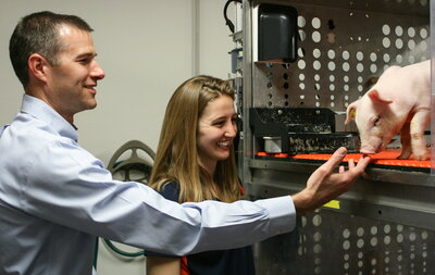
In the study, the research team brought 2-day-old piglets to their facility, known as the Piglet Nutrition and Cognition Laboratory, which is outfitted with large individual enclosures that let pigs see, smell, and hear others in adjacent pens. Dilger says the high ambient temperature and ability for pigs to socialize is important.
“In our nutritional studies, we want to keep them separated to avoid cross-contamination of the bacteria found in their colon, which is collectively known as the microbiota. But the pigs still get to express social behaviors by seeing, hearing, and smelling each other,” he says. “And in many of our studies, we let the pigs out of their enclosures to socialize each day, so they get to have a piggy party each afternoon.”
Another subset of piglets stayed with their littermates and mothers in farrowing crates on a research farm on the U of I campus. At 4 weeks of age, when pigs have developed enough that they no longer need to solely drink milk, the artificially reared pigs moved back to the farm and were group-housed with their sow-reared counterparts. So, in the end, the pigs only lived in different environments for the first four weeks of life and from that point forward, all pigs were treated the same.
All pigs were anesthetized and scanned in a state-of-the-art magnetic resonance imaging (MRI) machine at 1, 2, 3, 4, 8, 12, 18, and 24 weeks of age. The researchers assessed brain macro- and microstructure of the artificially reared and sow-reared pigs using the new pig brain atlases for young and adolescent pigs.
Not only did the pigs eat and grow at the same rates in the two rearing environments, their brain development was equivalent overall as well. The researchers found no differences in absolute volumes of the whole brain, gray matter, white matter, cerebrospinal fluid growth, or microstructural changes (neuronal connections between brain regions) over time in the two groups.
Joanne Fil, doctoral student in the Neuroscience Program at Illinois and first author of the study, says, “We looked at a surrogate measure of myelin, myelin water fraction, which reflects the fat and protein surrounding neurons and helps them communicate more effectively. Humans and pigs develop a lot of myelin significantly after birth, so if we see more myelination, then we assume the brain is maturing at a different rate. Longitudinally, there were no differences between the myelin water fraction in the two groups.
“We did see slight differences in the rate of development, with artificially reared pigs having a slightly higher rate of myelin development than the sow-reared pigs, but in the end, pigs raised in either environment reached the same place when it came to brain growth.”
The researchers also compared the pigs’ memory in behavioral tests. The pigs were presented with two stationary toys to play with – one they had previously been able to investigate and one they had never seen before. If they spent more time checking out the new toy, that was evidence they remembered the older one and therefore had encoded a memory.
At a couple of early time points, 4 and 8 weeks, pigs in the sow-reared group had slightly greater object recognition. The authors suggest that might have been due to greater peer interaction in the sow-reared environments. However, the differences were slight and temporary.
“Behavior is always more variable, more subjective. That’s why we like the objective and structural measures of the brain we can evaluate by MRI,” Dilger says. “And what we found is that when both sets of pigs are healthy and we've met their nutritional requirements, the rearing environment didn’t appear to influence brain development. Their brains effectively grew the same.”
In addition to supporting the continued use of laboratory rearing environments in pig neuroscience research, the study provides new data on pig brain development over time.
For half a dozen years, the first pig brain atlas served as the definitive reference for researchers around the world. But that atlas was based on 4-week-old pigs and couldn’t be easily extrapolated to older animals. Not only do the new atlases include one for 12-week-old pigs, the rearing environment study provides absolute volume measures for pigs at many more time points, up to adulthood.
“This provides a lot of foundational data where somebody could come back and ask how many cubic millimeters is the pig brain at a particular age. We can model that based on what we have here,” Fil says. “Our objective was comparing the two rearing environments, but also providing what is expected for a certain age of pig. That helps because we can start to develop nutritional interventions for specific ages and understand what parts of brain development can be influenced by nutrition.”
Dilger adds, “There's a lot of power in the pig as a model for biomedical research, and we’re showing that by bringing together engineering and agriculture, which are central to our mission as a land-grant institution. We use lots of mice in research on our campus, but we're also very good at working with pigs. We’re mixing the biomedical world with the agricultural world, to ultimately benefit both pig and human nutrition.
The article, “Influence of rearing environment on longitudinal brain development, object recognition memory, and exploratory behaviors in the domestic pig (Sus scrofa),” is published in Frontiers in Neuroscience [DOI: 10.3389/fnins.2021.649536]. Authors include Joanne Fil, Sangyun Joung, Courtney Hayes, and Ryan Dilger. Funding was provided by Nestlé.
The Department of Animal Sciences is in the College of Agricultural, Consumer and Environmental Sciences at the University of Illinois.
Sources: Ryan Dilger, 217-333-2006, rdilger2@illinois.edu;
Joanne Fil, jfil2@illinois.edu;
News writer: Lauren Quinn, 217-300-2435, ldquinn@illinois.edu
Date: April 14, 2021
Editor’s note: Images to accompany this story are available at https://uofi.box.com/s/s5g0mhioc8tkqt3hj8vkscyd0hw70gwk