Information about the Ko Laboratory.

Body

Lab Group Photo
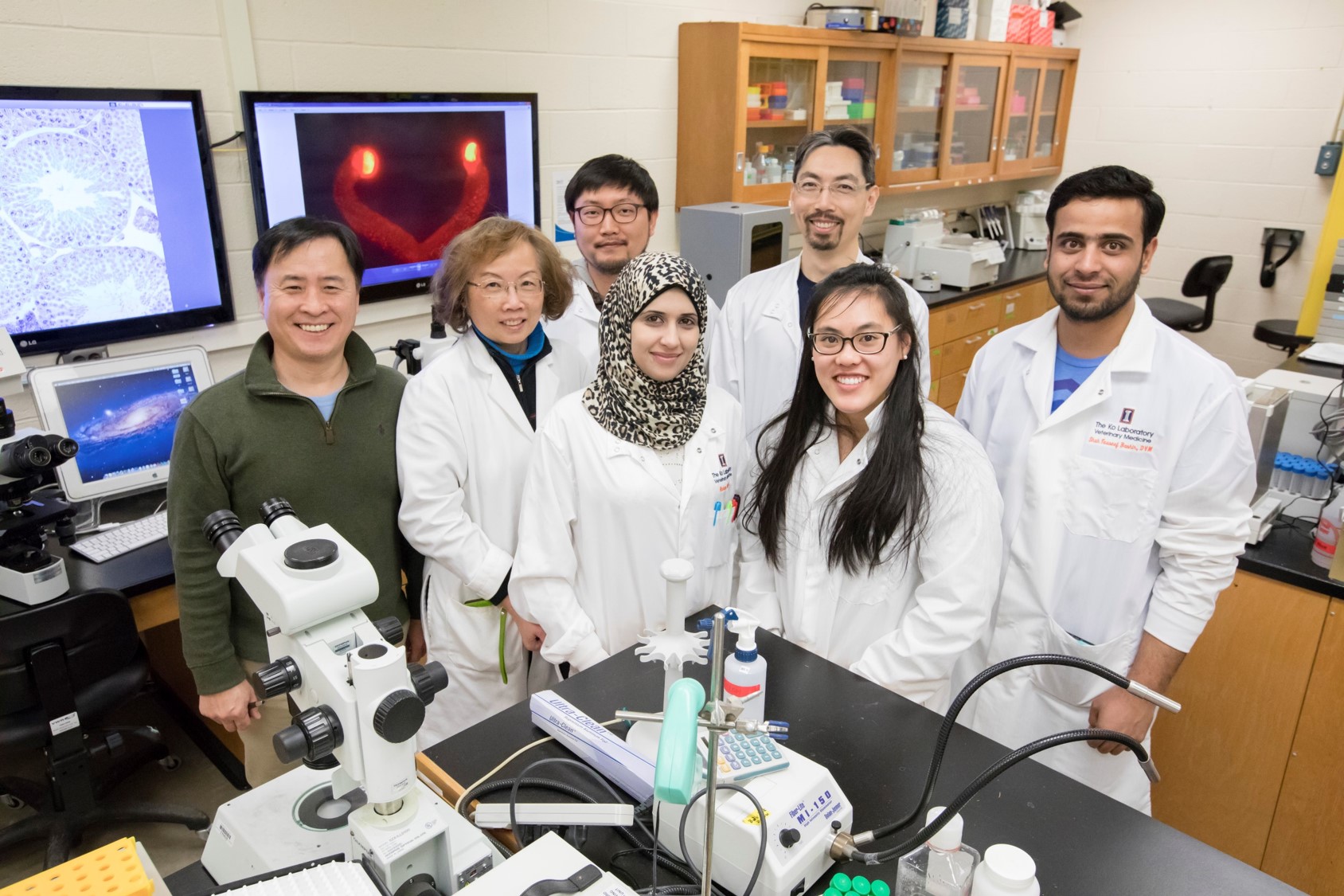
Thank you for visiting the Ko Laboratory page, a place for junior scientists who have a passion for science. This is their laboratory to achieve their goals for learning scientific skills, experimenting with new ideas, growing to excellency, and developing new knowledge for all. These goals are achieved by working on a few projects.

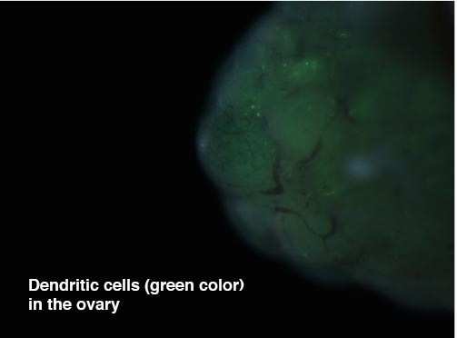
One of the main themes of the laboratory is to elucidate the mechanism governing the breakdown and rupture of the follicle during the ovulatory process. This process is activated by a surge of luteinizing hormone (LH), a pituitary gonadotropin, which triggers dramatic changes in the ovary at molecular, biochemical, and physical levels, eventually leading to the rupture of a mature follicle, releasing an egg. However, many of the key factors involved in and the mechanism governing the process of LH release and follicle rupture are yet to be unveiled.

Our research team aims to reveal the LH-induced intraovarian molecular events and the roles that ovarian steroid hormones play in this process, ultimately to help women who suffer from infertility caused by ovulatory defects. Other exciting projects include reproductive toxicology, novel roles that sex steroids play in reproductive/non-reproductive tissues and generation of novel transgenic animals and databases for shared uses.

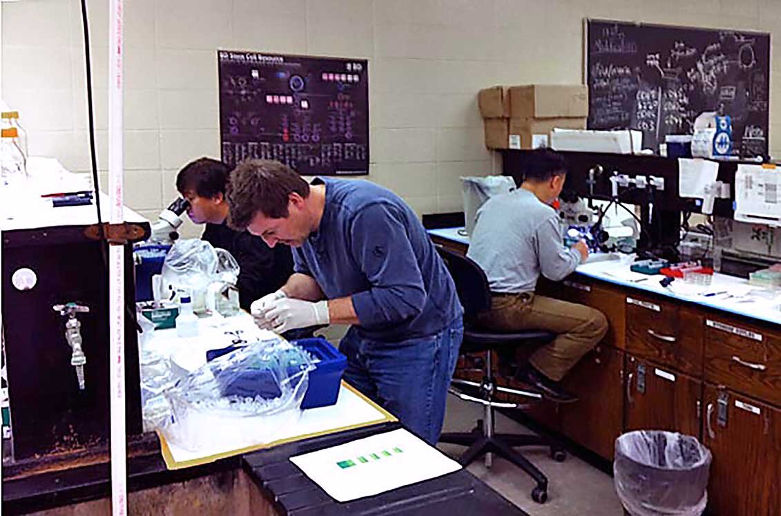
Please take a moment to explore our projects in the tabs below or see some funny or serious photos taken by our team members in the gallery below.

Our Principal Investigator is CheMyong Ko.

Please contact any of our team members if you are interested in our projects or want to collaborate with us. You can find their email contacts and projects at our website here.

Title
Projects
Endothelin-2 in ovulation and luteal formation

Endothelin-2, a short 21 amino-acid peptide, is a product of the highly conserved Edn2 gene that we hypothesize to be involved in both ovulation and angiogenesis of the corpus luteum. It was originally identified in porcine aortic endothelial cells as a potent vasoconstrictor, one of the most potent discovered. In the last 25 years, Endothelin-2 is expressed in a variety of tissues. While attempting to identify new genes that might be critical for ovulation using a microarray exploration and subsequent protein analysis, Endothelin-2 was found to be quite highly expressed in the granulosa cells of rodent ovaries at the time of ovulation. Endothelin-2 mRNA expression spikes for two hours just before the time ovulation, before returning to the essentially nonexistent basal levels that are otherwise present throughout the ovary, regardless of cycle.

As a potent vasoconstrictive peptide, Endothelin-2 has been hypothesized to act on myoid cells in the ovary that are present around the granulosa cells of mature follicles. Contraction caused by Endothelin-2 could be a direct final trigger for ovulation. Supporting this, ovaries treated with Endothelin-2 strongly contract ex vivo, while those treated with Endothelin Receptor antagonist drugs ovulate fewer oocytes. As an ongoing follow up approach, we are now utilizing a transgenic mouse model in which Endothelin Receptors are absent only from the smooth muscle cells of the ovary, in the expectation that these mice will have difficulty ovulating, allowing the effect of Endothelin-2 to be pinpointed to those contractile cells

The Ko Laboratory specializes in transgenic animal models, and a second approach to look at the role of Endothelin-2 in ovulation using a mouse that globally lacked the Edn2 gene expression was also used. As expected, these mice ovulated fewer oocytes than control wild type mice, highlighting the overall importance of Endothelin-2. Interestingly, these mice all also lacked corpora lutea even if they successfully ovulated. The corpus luteum is a highly vascular structure that forms after ovulation and is necessary for progesterone synthesis and pregnancy maintenance. We are now focusing on generating mice that lack the Edn2 gene only in the ovary, under the belief that Edn2 is necessary for vessel formation through VEGF induction, endothelial cell expansion, MMP secretion, and/or monocyte invasion. We hope that this exploration can eventually be applied to either improve human and food animal fertility, or in the generation of novel contraceptive approaches.

Recent publication: Granulosa cell endothelin-2 expression is fundamental for ovulatory follicle rupture

Progesterone receptor in regulating Kisspeptin secretion

A subset of hypothalamic neurons releases a neuropeptide that stimulates GnRH neurons to secrete GnRH. This molecule, Kisspeptin, is a 10-amino acid long small peptide synthesized by a post-translational cleavage of a pro-peptide. Kisspeptin neurons are localized to two hypothalamic nuclei: anteroventral paraventricular nucleus (AVPV) and acute nucleus (ARC). Estrogens via its receptor, estrogen receptor alpha (ERα or ESR1), positively regulate Kisspeptin secretion in the AVPV but negatively in the ARC. Upon stimulation by Kisspeptin, GnRH neurons release GnRH that in turn stimulates the pituitary gonadotrophs to secrete follicle-stimulating hormone (FSH) and luteinizing hormone (LH) leading to ovarian follicle growth and ovulation.

Progesterone receptor (PGR) is a nuclear receptor transcription factor. In the ovary, PGR plays a critical role in ovulation and corpus luteum (CL) formation.  Formation of CL is necessary to support pregnancy by progesterone production by the newly formed CL. Progesterone also positively and negatively regulates the estrous cycle, likely through interactions with its receptor. In the hypothalamus, PGR is expressed in the Kisspeptin neurons in both the AVPV and ARC and its expression levels fluctuate during the estrous cycle.

We are investigating the functional role that PGR plays in kisspeptin neurons. We hypothesize that PGR plays a critical role in releasing Kisspeptin. To facilitate the investigation, our laboratory developed a unique mouse model in which Pgr is deleted specifically in the Kisspeptin neurons. We find that the conditional Pgr knockout mice are born fertile, but lose fertility prematurely. In support of our hypothesis, these mice display a significantly lower basal serum LH level, indicating defective GnRH secretion presumably caused by reduced stimulation by its upstream regulation, Kisspeptin.

Recent publication: Loss of Fertility in the Absence of Progesterone Receptor Expression in Kisspeptin Neurons of Female Mice

Regulation of leukocyte infiltration into the ovary

In women of reproductive age, cross-talk between the immune and reproductive systems impacts fertility. In particular, studies have shown that tight spatiotemporal control of leukocyte infiltration into the ovary is pivotal for successful ovulation. Once infiltrated, the leukocytes differentiate into a cohort of pro- or anti-inflammatory cells in the preovulatory ovary and play critical roles in ovulation and luteal formation. Our laboratory investigates the mechanism of leukocyte infiltration and the role of progesterone (P4) and prostaglandins (PGs) in the process. This study aims to identify mediators of P4 and PG action in regulating ovarian WBC infiltration and to identify the trigger of preovulatory splenic WBC release. This will lead to better diagnoses and more efficient clinical treatment for defects in ovulatory function.

Recent publication: Pituitary-ovary-spleen axis in ovulation

Generation of transgenic animal models

mice reproductive organs and brains
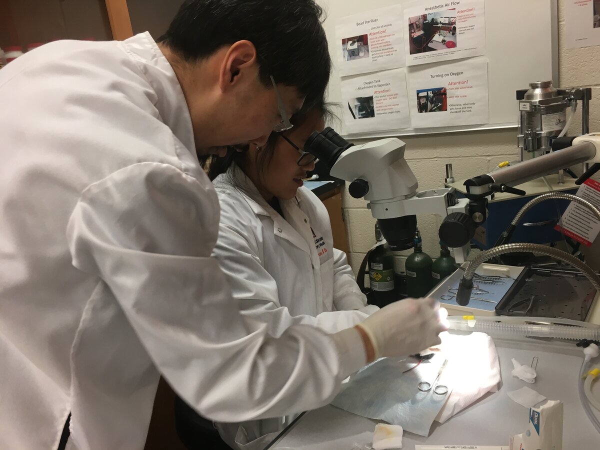
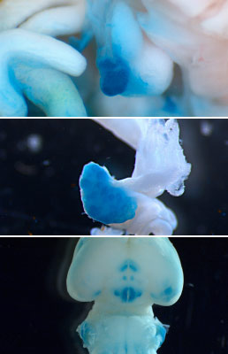
Our lab generates genetically modified (or transgenic) animal models. Genomic DNA sequences are modified using a variety of genetic manipulation procedures in vitro. The DNA sequences are then introduced into the genomes of recipient animals, creating new animals that never existed before. New animal models are continuously generated and used in our laboratory.

Shown are the microscopic images taken from the newly generated transgenic mouse in which estrogen response tissues automatically labeled in blue color. Displayed to the right are the male reproductive organs (top panel), female reproductive organs (middle) and ventral side of the male brain (bottom).

The following lines of transgenic animals were developed by our research team:

Cyp17-iCre: This is the first and most widely used transgenic mouse line among those created by our laboratory.  This mouse is used for conditional deletion of genes in the androgen-producing cells and also for localizing cells that synthesize androgens.  The generation and characterization of this line of mice were published in Genesis in 2008. This line is now available via JaxMice.

End2-iCre: This mouse line is used for conditional deletion of genes in endothelin-2 expressing cells and for localizing the sites of endothelin-2 expression.  The generation of this line of mice was published in February 2015 as a cover image of the journal Genesis.  There is a high demand for this line of mice from a variety of disciplines of science.  This line is now available via JaxMice.

Edn2-flox: This mouse line is used for conditional deletion of endothelin-2 gene.  We used this line of mice for demonstrating the critical role that endothelin-2 plays in ovulation (Scientific Reports, 2017).  Endothelin-2 is also expressed in the eyes, intestines, and kidneys. This line of mice is already being requested by other laboratories.

Esr2-iCre: This mouse line is used for conditional deletion of genes in the ERb (estrogen receptor beta)-expressing cells.  This line is also used for localizing the sites of ERb expression.  The generation of this line was published in Genesis in 2016.  A grant was submitted to NIH for the use of this model in studying estrogen receptors in the ovary. This line is now available via JaxMice.

Esr1-iCre: This mouse line is used for conditional deletion of genes in the ERa (estrogen receptor alpha)-expressing cells.  This line is also used for localizing the sites of ERa expression.  The generation of this line is yet to be reported as a manuscript.

Recent publications:
Generation of an estrogen receptor beta-iCre knock-in mouse
Generation and characterization of an endothelin-2 iCre mouse

Ovary in female aging

Aging female ovaryFemale reproductive senescence begins before birth and continues throughout a lifetime, culminating with menopause, the state of depletion of ovarian follicles. The female reproductive axis is unique in that it reaches a senescent state when other organs in the body are generally healthy. On average, women reach menopause at 50 years of age whereas their life expectancy is over 80 years. Hence, the majority of women will spend, or are spending, more than one-third of their life in a post-menopausal state. Epidemiological studies show that late onset of menopause confers longevity and decelerates the appearance of much age-related morbidity, suggesting that delaying the onset of menopause will improve the health of aging women and therefore their quality of life. In this study, using animal models, we aim to develop an experimental procedure that will be conceptually applicable to delaying the natural onset of menopause in women. Once developed and successfully executed, this novel procedure may delay the onset of menopause by multiple years so that women may not experience menopause until they reach the age of 55, 60 or greater.

Our project is based on the idea that by preserving ovarian tissues (cortices where most primordial follicles reside) at a young age when follicles are densely populated and then grafting this tissue back to her own ovary at an old age when follicles are sparsely populated, we will be able to increase the recipient’s follicle pool size. The increased follicular pool may, therefore, extend the functional life span of the ovary, resulting in a delay of menopausal onset. In this project, we aim to simulate human ovarian tissue grafting using well-established mouse menopausal models: naturally aging mice and 4-vinylcyclohexene diepoxide (VCD) injected mice. Briefly, young ovarian cortices will be transplanted to an older ovary, and we will determine whether the transplantation delays the onset of menopause of the recipient. Specifically, we will determine the effect of transplanting post-pubertal ovarian tissues to pre-, peri- and post-menopausal animals and measure the on the onset of menopause.

Our laboratory has successfully established an ovarian tissue transplantation protocol using the C57BL/6 strain of mice. Some studies in rodent models, however, have shown that the hypothalamus of a middle-aged female (when the ovary is functional) is less sensitive to preovulatory estradiol positive feedback stimulation, resulting in a reduction in the number of activated GnRH neurons and a delayed or attenuated secretion of LH from the pituitary. These observations may indicate that hypothalamic senescence may precede ovarian aging with the possibility that ovarian cortices may go through accelerated aging if transplanted to an older recipient. However, other studies suggest that ovarian aging itself is the predominant factor for the aging of the reproductive axis and, therefore, the onset of menopause.

In this study, we will use our ovary transplantation procedure to resolve these conflicting reports by measuring the aging rate of ovarian cortices when transplanted to different age groups of mice and the aging rate of the hypothalamus when varying age groups of mice receive ovarian cortices from different age groups of mice.

Shown at the top right is a microscopic image taken from an ovary that received an ovarian tissue graft that was taken from a green fluorescent mouse.

Progesterone receptor in regulating ovarian follicle rupture and inflammation

LH triggering before and after
LH trigger: Before and After

Luteinizing hormone (LH) is secreted from the pituitary and triggers ovulation. Progesterone is synthesized in the ovary upon stimulated by LH, and activates progesterone receptor (PGR) leading to ovulation. Meanwhile, LH also induces the expression of prostaglandin-endoperoxide synthase 2 (Ptgs2) gene, then granulosa cells produce the inflammation trigger, prostaglandins via enzymatic activity of PTGS2. To understand the role of PGR in ovulation and ovulatory inflammation, we are utilizing molecular biological tools and genetically modified animal models including granulosa cell-specific Pgr knockdown mouse (Esr2-PgrKO).

 RecentlyChart of PTGS2 Expression 
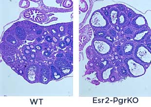
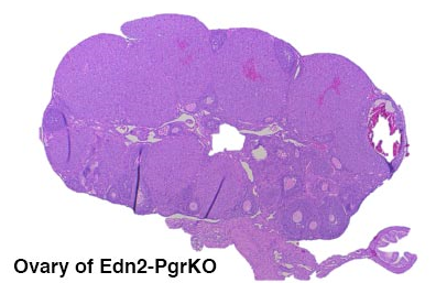
we found that PTGS2 expression was potentially attenuated by PGR in the granulosa cells. Therefore, we hypothesize that PGR activation suppresses cytokine productions, leukocyte recruiting and ovulatory inflammation via inhibiting PTGS2 expression. We will identify the inhibition mechanism of PGR on PTGS2 expression and anti-inflammatory role of PGR in the preovulatory ovary.

Effect of DEHP on gonadal development

Mouse Fertility Chart

The main goal of this study is to determine the effects of environmental chemicals on gonadal and brain development in male mice. Phthalates are a family of synthetic chemicals that are widely used in medical, automotive and consumer products. Di-(2-ethylhexyl) phthalate (DEHP) is an endocrine disruptor that has anti-androgenic activity. Male CD-1 mice were prenatally exposed to DEHP by dosing their mother daily with 200 μg, 500 mg or 750 mg/kg or vehicle from gestation day 11 to birth. The impact of the exposure on their reproductive and memory was examined when they reached the ages of 8 to 22 months. As early as at the ages of 8 months, the 750 mg/kg/day DEHP group of mice exhibited significantly reduced fertility while other dosage groups and control mice did not. The same dose-dependent reduction of fertility continued until the age of 18 months.

All DEHP-exposed group had lower serum testosterone and higher serum estradiol levels than the control mice. Histological evaluations showed that mice prenatally exposed to DEHP displayed a wide array of gonadal and epididymal abnormalities such as increased germ cell apoptosis, degenerative seminiferous tubules, oligozoospermia, asthenozoospermia, and teratozoospermia in comparison to age-matching control mice. Behavioral tests were performed at the ages of 16-17 months to examine the memory and anxiety level. Elevated plus maze test and open field test showed an increase in the anxiety behavior in DEHP treated mice compared to control. Novel object recognition test and Y-maze test found a significantly reduced recognition memory and spatial memory in DEHP-exposed mice. In addition, we will determine whether the potential impact of the exposure is transmitted to the next generations via epigenetic passages. This study will eventually help to develop a preventive measure to protect children from maternal exposure to environmental toxicants.


Recent publication: Prenatal Exposure to DEHP Induces Premature Reproductive Senescence in Male Mice

Endothelin-2 in hair follicles

The purpose of this project is to investigate the function of EDN2 in the epidermis, specifically its potential role in the maintenance of melanocyte stem cells within hair follicles. We will examine the spatial and temporal expression of EDN2 in the epidermis of mice via fluorescent microscopy using a novel transgenic mouse (edn2-GFP) and other relevant methods. We will also conduct microsurgeries involving skin transplantation, in which skin tissues from edn2 knockout mice would be grafted onto wild-type mice, so as to observe how the loss of EDN2 expression would impact hair/skin growth/pigmentation over a longer period of time.

Progesterone receptor in the ovary

dendritic in ovary

Previous studies have revealed that the hormone progesterone, along with several types of immune cells, play a pivotal role in ovulation. Dendritic cells are one such type of immune cells that act as ‘messenger cells’ within the body’s immune system. While it is known that the presence of both dendritic cells and progesterone is necessary for ovulation to occur, the correlation between them is not fully clear. This project focuses on understanding this mutual interaction between progesterone and dendritic cells, and how they impact each other to make ovulation possible. We are particularly interested in studying how dendritic cell numbers vary depending on the expression of progesterone within the ovary. We are comparing levels of dendritic cells within the reproductive tracts of female mice, in the presence and absence of progesterone, using Yellow Florescence Protein (YFP) as an identification marker. Hence, the study will enable us to further understand the coordination between dendritic cells and progesterone, the specific role of dendritic cells with regards to ovulation, and how the dynamic process of ovulation ultimately takes place.


Progesterone receptor in the corpus luteum

Ovary of Edn2-PgroKOThe focus of this study is to determine the functional role of progesterone receptor in regulating progesterone synthesis in the Corpus Luteum. We hypothesize that progesterone receptor is required for continued synthesis of progesterone during the luteal phase. For this study, we use transgenic mice that are selectively deficient of progesterone receptor in the luteal cells. To confirm the functioning role of the progesterone receptor, fertility, P4 concentration in the serum and the ovary are being currently assessed. Future studies include luteal cell cultures and determination of steroidogenic gene expression under the deficiency of progesterone receptor.

Recent publication: Beclin-1 deficiency in the murine ovary results in the reduction of progesterone production to promote preterm labor

Conversion of an Esr1+ cell to Esr2+ cell during ovarian development

Locationization of ESRs in the ovary
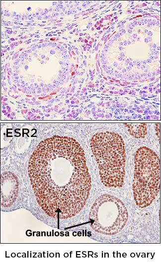
17β-estradiol (E2) is a steroid hormone that regulates a plethora of reproductive, metabolic, immunologic, cognitive, and skeletal functions. Two classical estrogen receptors, ESR1 (ERα) and ESR2 (ERβ) are responsible for the classical actions of E2 in mammalian species. In the ovary, ESR1 is localized to the surface epithelium and theca cells, whereas ESR2 is expressed in the granulosa cells.

In previous studies, we have generated a transgenic mouse line that has an insertion of an enhanced Cre (iCre) recombinase in the exons of Esr1 and Esr2 gene, respectively. Esr1Cre mice can be used for deleting any floxed gene in the ESR1-expressing (Esr1+) cells, and Esr2Cre mice for excising genes in ESR2-expressing (Esr2+) cells. These novel transgenic mice are being used for lineage-tracing Esr1+ and Esr2+ cells and investigating unique roles that these estrogen receptors play in a single lineage. One interesting finding that we made is that Esr2+ granulosa cell seems to originate from cells that once expressed ESR1. We hypothesize that there is a molecular switch that converts Esr1+ cells to Esr2+ cells in developing ovary.

In this project, we have three main goals:

  1. Determining the temporal window when ESR1/ESR2 switching occurs
  2. Determining the role of oocytes in granulosa cell lineage determination
  3. Discovering the mechanism of silencing Esr1 promoter during the transition of ESR1 to ESR2 granulosa cells.

The discovery of the existence of a mechanism that converts ERa+ cells to ERb+ cells is expected to open up a possibility of changing Esr1+ cells to Esr2+ cells.

Investigation of ESR2 role in the cecal development and function

Estrogen has been well known to have protective effects in various diseases to a certain degree. However, estrogen via estrogen receptors (ERa and ERb), may have an essential but unidentified role in preventing certain intestinal diseases like colorectal cancer (CRC). Colorectal cancer is the fourth most common cancer and is diagnosed more in men compared to women. Studies have shown an association with increased tumor size and cell proliferation in patients diagnosed with CRC and decreased ERb. ERb, the predominant receptor in the colon and cecum, has anti-proliferative effects. Many individuals diagnosed with ulcerative colitis advance to CRC. The goal is to target and understand ERb and its effects on ulcerative colitis before a more severe condition like CRC.

The objective of this project is to:

  1. determine the spatiotemporal expression of ERb in the developing and adult ceca
  2. determine the impact of ERb-deficiency on cecal development
  3. assess the effects of estrogen deficiency on cecal development
  4. determine the sex-dependent phenotype, and
  5. determine the impact of ERb-deficiency on colitis and the microbiome.

Currently, we are determining whether or not oxazolone-induced colitis is a good model for humans diagnosed with ulcerative colitis. After finding a good model for colitis, we will determine the impact of ERb-deficiency in mice with colitis.

Investigation of DEHP exposure on colonic inflammation

DEHP is a ubiquitous plasticizer contained in, but not limited to, PVC plastics, cosmetics, personal care products, gastrointestinal medications, and medical devices. This specific phthalate is noncovalently bound to plastics, and therefore, can easily leach into the environment and contaminate food and water sources. Many studies have examined the impact and toxic effects of DEHP exposure on reproductive, cardiovascular, hepatic, cognitive, renal, and respiratory health. However, no studies have examined the effects of DEHP exposure on the gastrointestinal tract, specifically the colon, and this is critical to research because 1) human exposure to DEHP is mainly by ingestion and 2) the symptoms of inflammatory bowel disease may be exacerbated due to DEHP exposure.

Our study will be the first to determine the innate and adaptive immune responses of environmentally relevant doses of DEHP exposure. Future objectives of this project are to:

  1. Determine the effects of DEHP exposure on the colonic microbiome and how it may mediate intestinal immune responses
  2. Assess the impact of DEHP exposure in mice induced with colitis
  3. Determine the sex-dependent phenotype