![What Causes Tumors? [Guide to Benign and Malignant Tumors]](https://vispdocs.com/wp-content/uploads/2021/08/tumor-cells.png)
The Legget Tumor Dynamics Lab applies bioengineering, live imaging, and computer vision to help diagnose and treat cancer. We focus on recreating realistic 3D environments that mimic the body, allowing us to investigate how cells behave in both healthy and diseased states, especially in different forms of cancers.
The goal of our lab is to advance personalized medicine by studying how individual patients’ cells react to treatments, helping create therapies tailored to each person’s unique biology.
Our Principal Investigator is Susan Leggett
For more info, visit our website!
Advanced imaging techniques
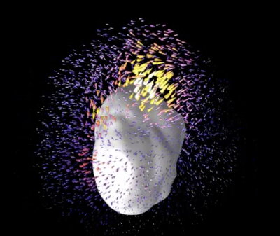
Using advanced imaging techniques, we rapidly produce detailed visualizations that reveal mechanical properties, such as stiffness, at both the cellular and tissue levels. These models simulate real-life conditions in the body, allowing us to observe how cells respond to their environment. This approach helps us investigate how dynamic cell behaviors and mechanical cues shape tissue function in both health and disease.
Organ-on-a-chip system
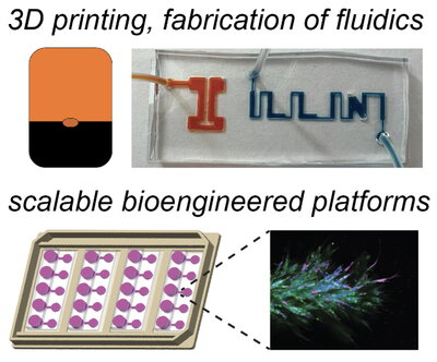
Our lab also utilizes 3D printing to recreate tissue microenvironments of interest. With our organ-on-a-chip system, we are able to replicate real-life conditions in the body and support live-cell imaging to observe how cells behave over time. Mechanical perturbations, tissue interfaces, and spatial heterogeneity are used to provide a controlled and measurable way to study how tissues are organized, how cells adapt, and how physical forces influence biology in both healthy and diseased states.
Reverse-engineering the breast and ovarian tissue
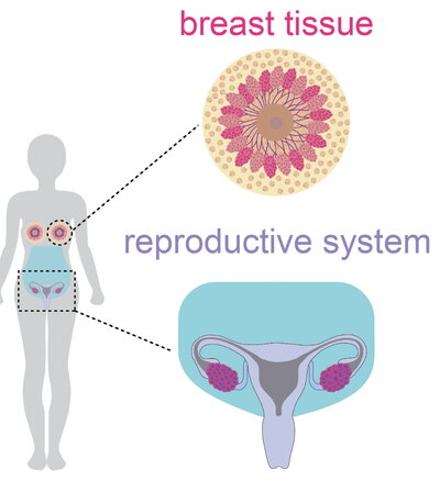
We have created tools to help reverse-engineer the breast and ovarian tissue using patient-derived samples. Using this tool helps us study cancer progression and therapeutic response in these areas. This helps model tumor dynamics for personalized medicine, as the patient samples allow us to study heterogeneity and treatment responses in a controlled setting.
Live-cell imaging
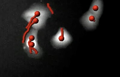
We perform live-cell imaging to study how cells behave, interact, and adapt. Using 3D environments, time-lapse fluorescence, and confocal microscopy, we can capture how cells move, grow, change, and spread over time and across different areas in the tissue